A specific lesion
Class I to V, depth, extent, pulpal involvement or not. The exact stage you want diagnosed or treated.
Mesial Class IICervical lesionDeep near-pulpal caries
Dental training models · Designed by clinicians · Made in Morocco
High-fidelity dental models designed by clinicians to make anatomy easier to understand, demonstrations clearer and clinical procedures easier to learn and practise repeatedly.
A blank molar, one instrument — and the movement to come.
Restorative collection
A structured range for observing, preparing, restoring, repeating and assessing clinical procedures.
01 · Restorative collection
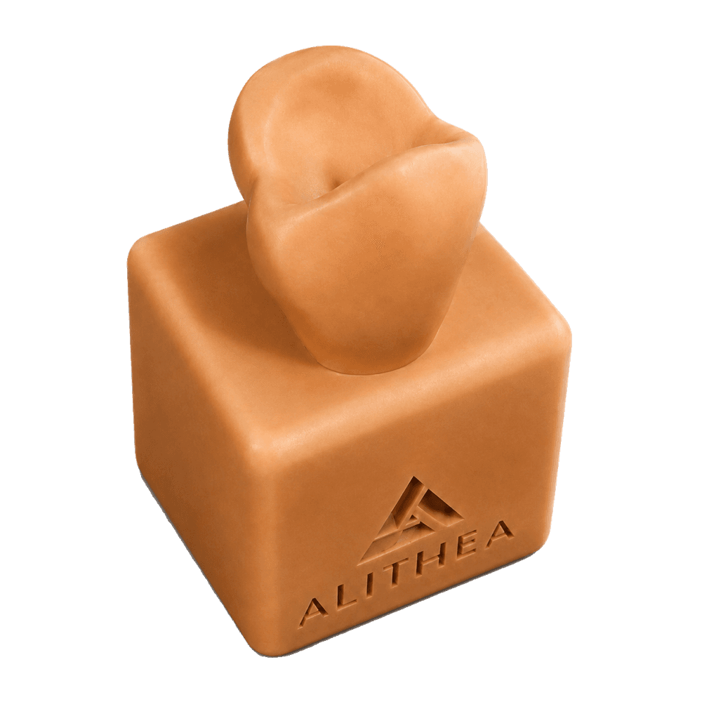
Single tooth. Infinite precision.
Single-tooth model: anatomical reference, morphology and wax-up training.
View the range02 · Restorative collection
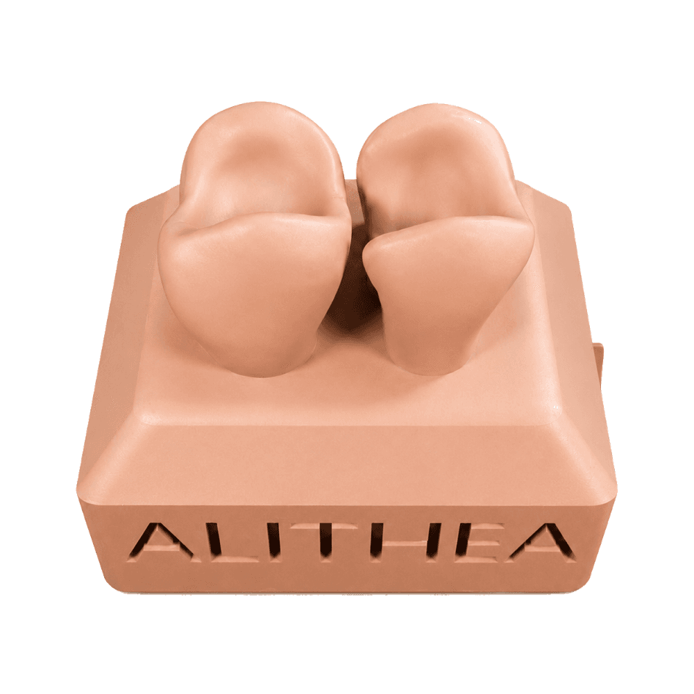
Two teeth. Mastered contact.
Twin teeth: Class II restoration and proximal contact management.
View the range03 · Restorative collection
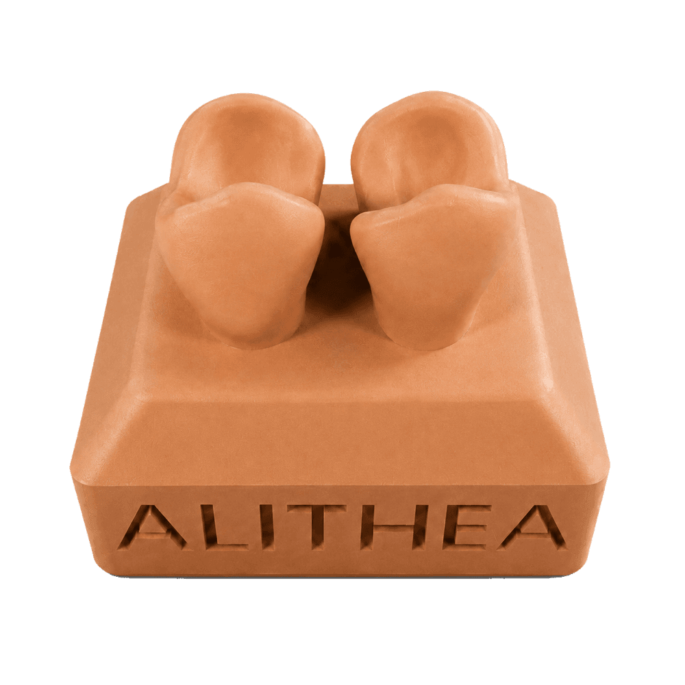
Two lesions. One gesture.
Mirrored proximal lesions: repeat the gesture twice, build muscle memory.
View the range04 · Restorative collection
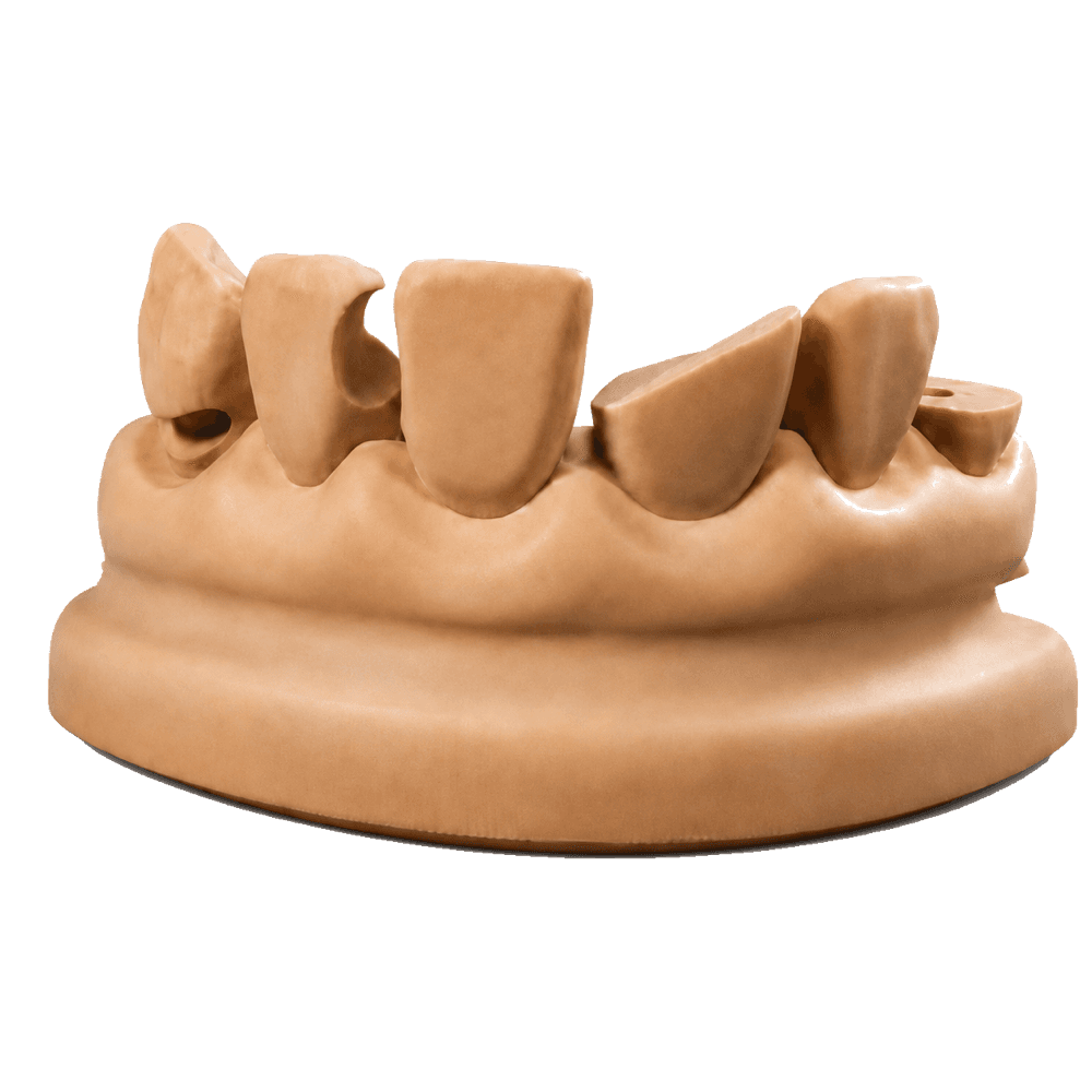
A spectrum of pathologies.
Full arch segment for multi-situation diagnostic training.
View the range05 · Restorative collection
The stamp. Anatomy restored.
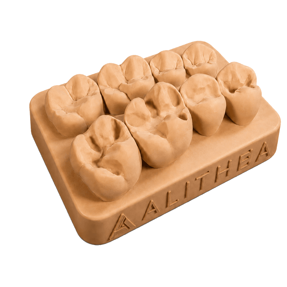
Preformed occlusal anatomy for learning and repeating the stamp technique: capture the original morphology, then rebuild it in composite.
View the rangeSurgical rehearsal
Send us your patient's CBCT and, if you have one, your surgical guide. We print a high-fidelity replica of their anatomy. Place your implants on it, check how the guide seats, verify angulation, depth and emergence — and go into surgery on familiar ground.
Submit a caseMandibule anatomique de référence — BodyParts3D, © Database Center for Life Science, CC BY-SA 2.1 JP. Aucune donnée patient.
Teaching design
You are a clinician, a teacher or a trainer, and the situation you want your students to work on is in no catalogue — yours or ours. Describe it: we draw it, you approve it, we print it.
What you can specify
Class I to V, depth, extent, pulpal involvement or not. The exact stage you want diagnosed or treated.
Complex extraction, impacted tooth, root separation, implant site. The difficulty you want rehearsed.
Atypical canal, curved root, malposition, rare morphology. What you meet once a year in the clinic.
The same case thirty times over, or a graded progression from one session to the next. Every student works the same thing.
No patient data required: these models are designed, not copied. A sketch, a textbook photo or three lines is enough to start.
Describe a caseHow our models are made
Every model is designed by clinicians with first-hand understanding of the clinical procedure it is intended to teach and help learners practise.
High-resolution additive manufacturing with carefully calibrated settings to reproduce fine anatomical detail.
Every component is cleaned, post-cured, inspected and assembled before shipping.
Protective packaging and dispatch from Casablanca. Delivery across Morocco and worldwide.
In development
Anatomical models developed for surgical education, planning and hands-on procedural training.
Root canal anatomies designed to make internal structures easier to understand and endodontic procedures easier to practise repeatedly.
In every parcel
Three decals drawn in-house, slipped into every shipment. For the scrubs, the locker, the instrument case.
Forma fidelis · Gestus certus
Three ways to order
Explore our collections or tell us about your educational project.