Atlas
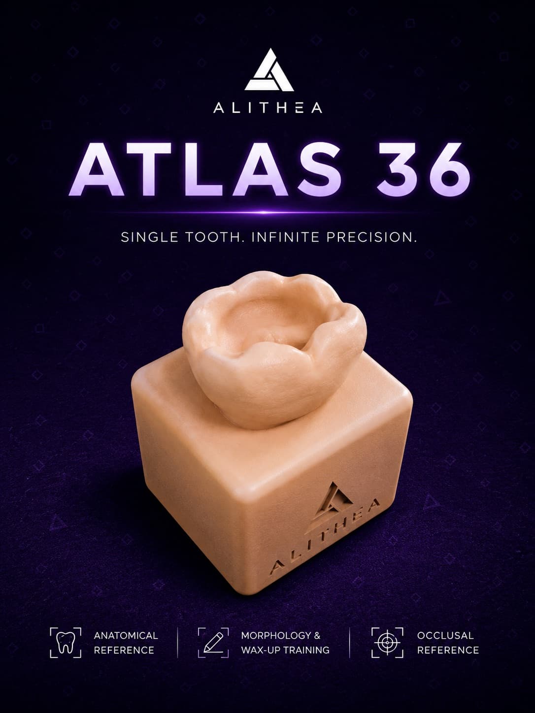
Atlas 36
A single-tooth anatomical model for studying occlusal landmarks, marginal ridges and proximal contacts.
MAD 14
Collection
Morphology, preparation and restoration on legible anatomical situations.
Single tooth. Infinite precision.
Single-tooth model: anatomical reference, morphology and wax-up training.
View range 02Two teeth. Mastered contact.
Twin teeth: Class II restoration and proximal contact management.
View range 03Two lesions. One gesture.
Mirrored proximal lesions: repeat the gesture twice, build muscle memory.
View range 04A spectrum of pathologies.
Full arch segment for multi-situation diagnostic training.
View range 05The stamp. Anatomy restored.
Preformed occlusal anatomy for learning and repeating the stamp technique: capture the original morphology, then rebuild it in composite.
View rangeAvailable models
Atlas
A single-tooth anatomical model for studying occlusal landmarks, marginal ridges and proximal contacts.
MAD 14
Atlas
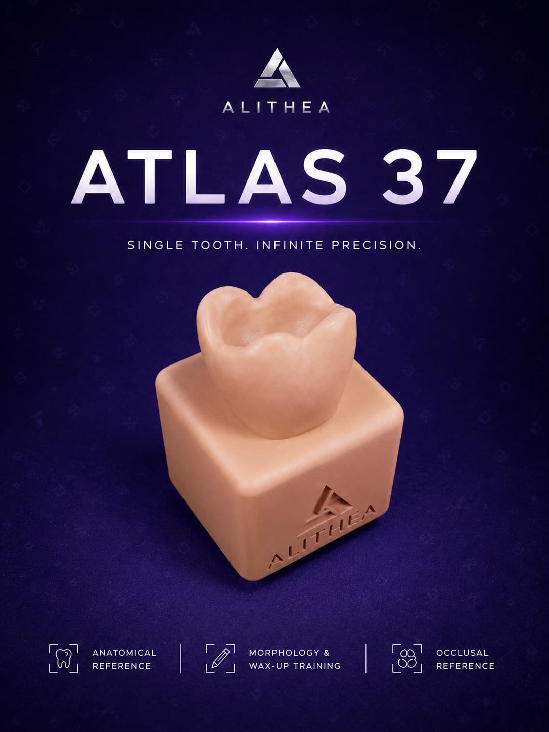
Enlarged anatomical reference of the lower left second molar.
MAD 14
Atlas
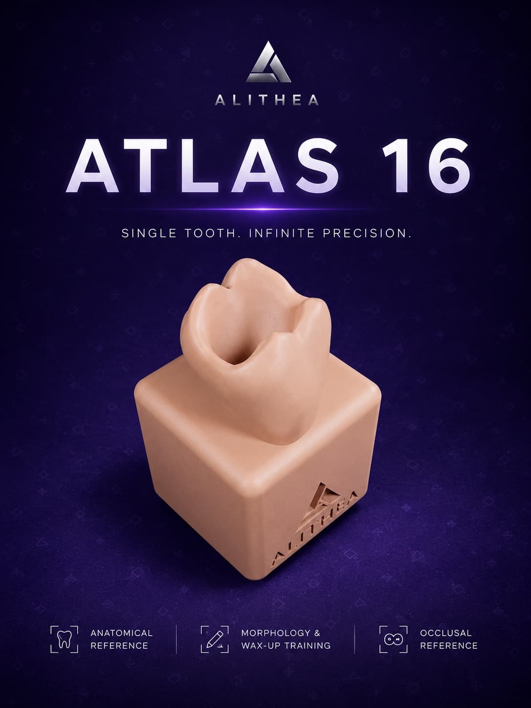
Three-rooted morphology of the upper first molar at teaching scale.
MAD 14
Atlas
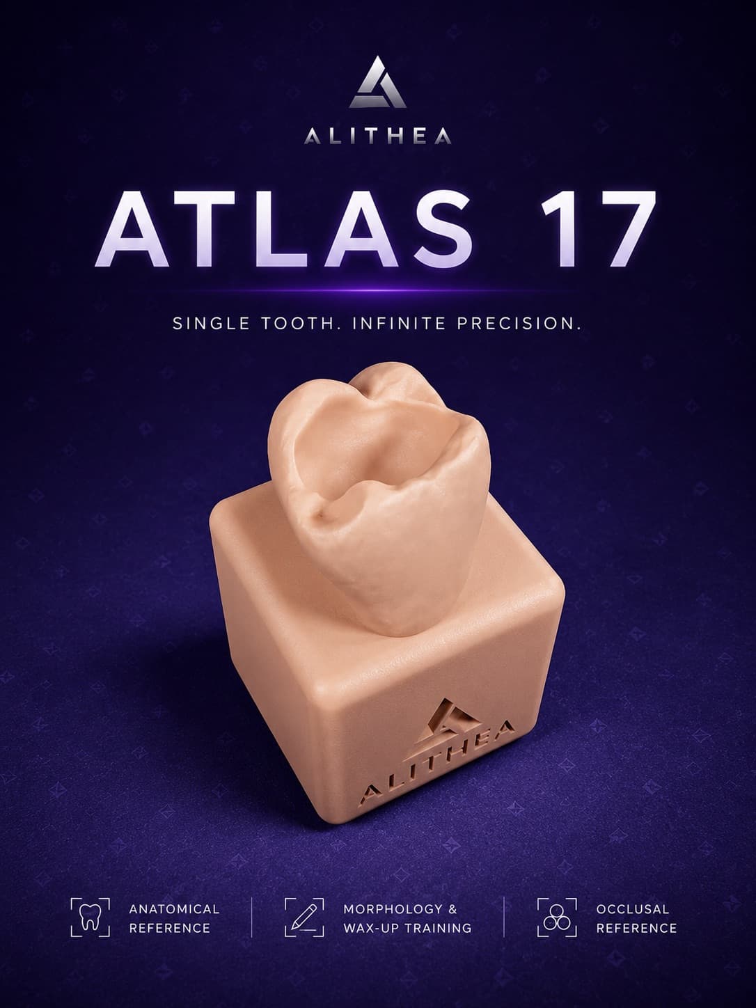
Enlarged upper second molar for inter-arch morphological comparison.
MAD 14
Atlas
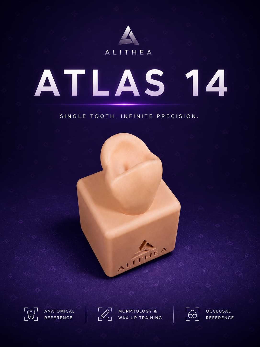
Upper first bicuspid at working anatomical scale.
MAD 14
Atlas
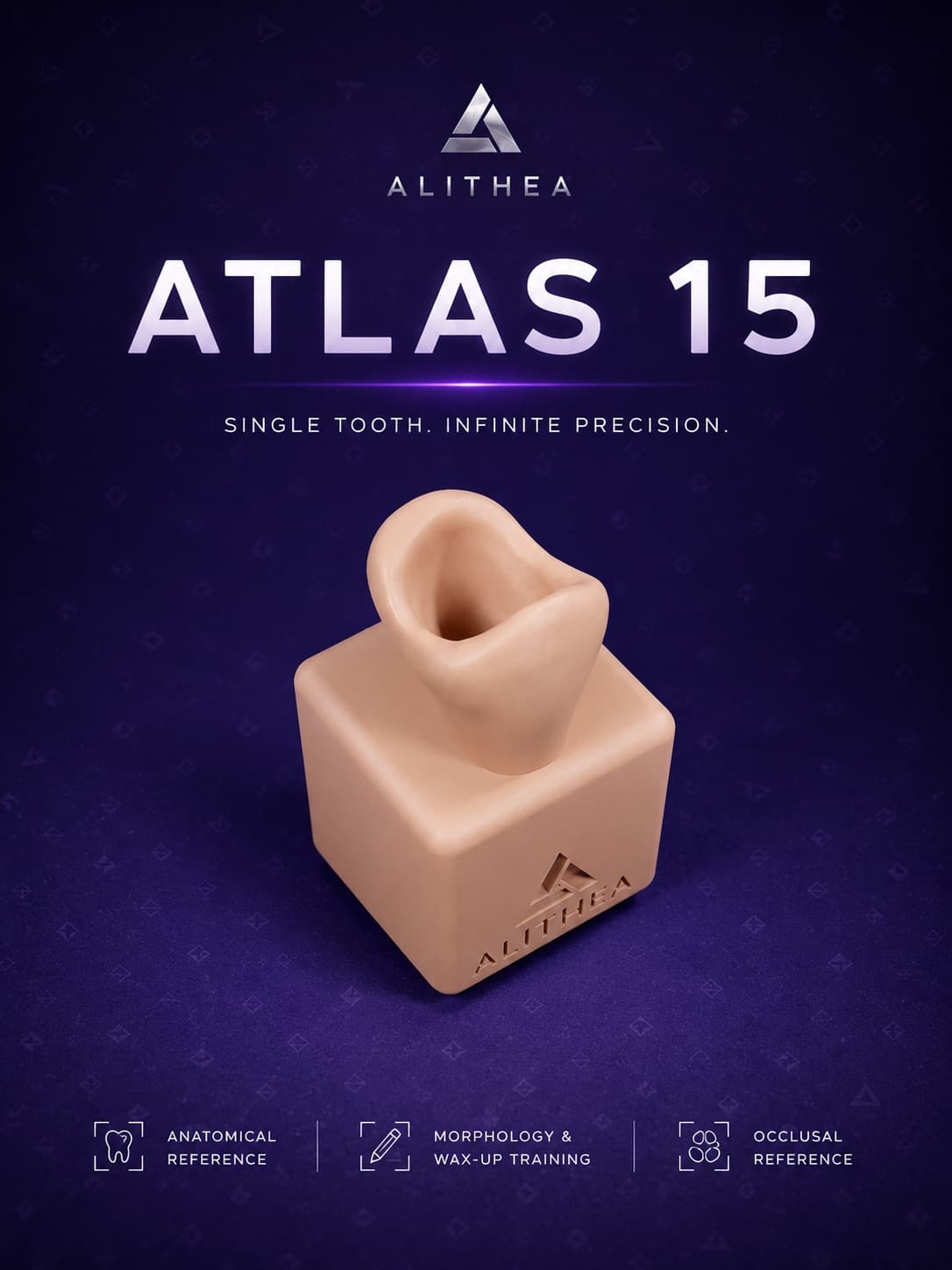
Upper second premolar for comparison and wax-up training.
MAD 14
Atlas
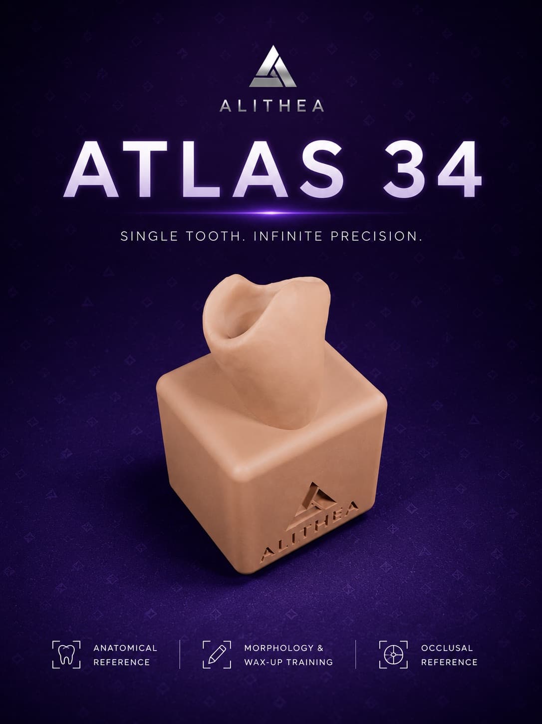
Functional lower monocuspid premolar at enlarged scale.
MAD 14
Atlas
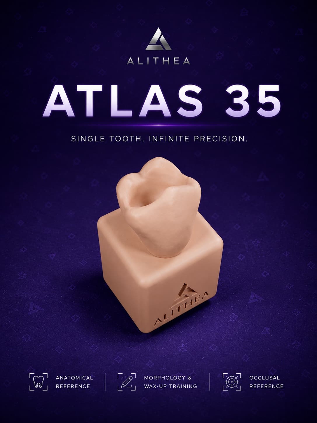
Second mandibular premolar with bicuspid morphology for advanced training.
MAD 14
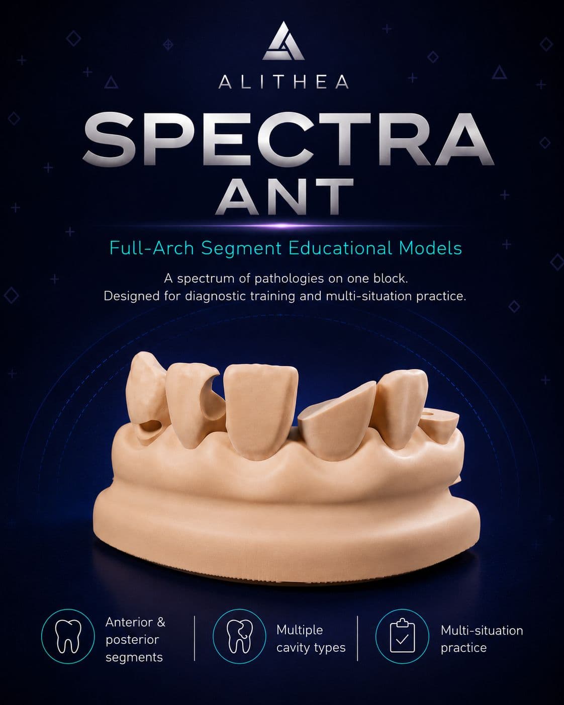
Spectra
A complete anterior arch segment, gathering several clinical situations on a single block.
MAD 34
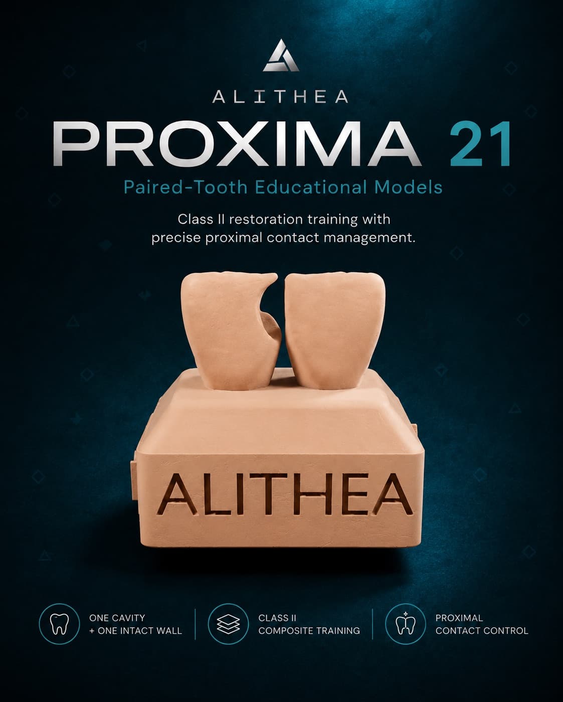
Proxima
Central incisors: a single proximal lesion on the 21, the 11 left intact as the symmetry reference.
MAD 20
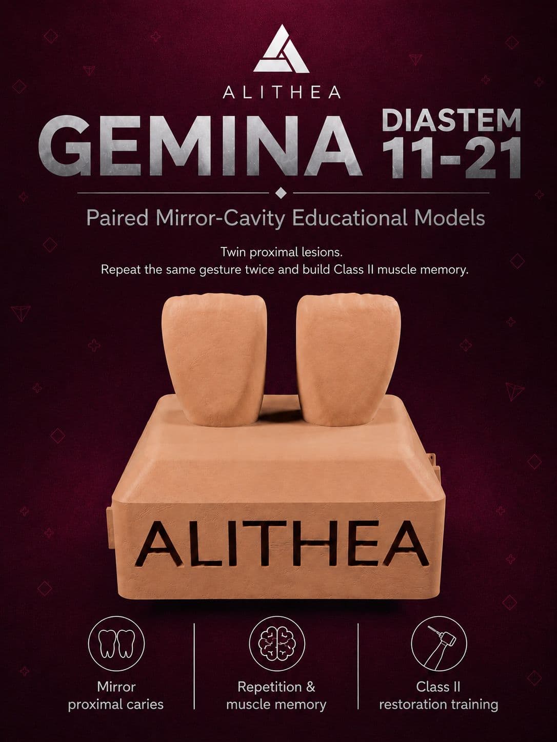
Gemina
Interincisal diastema closure: both centrals to be built up in mirror image with composite.
MAD 20
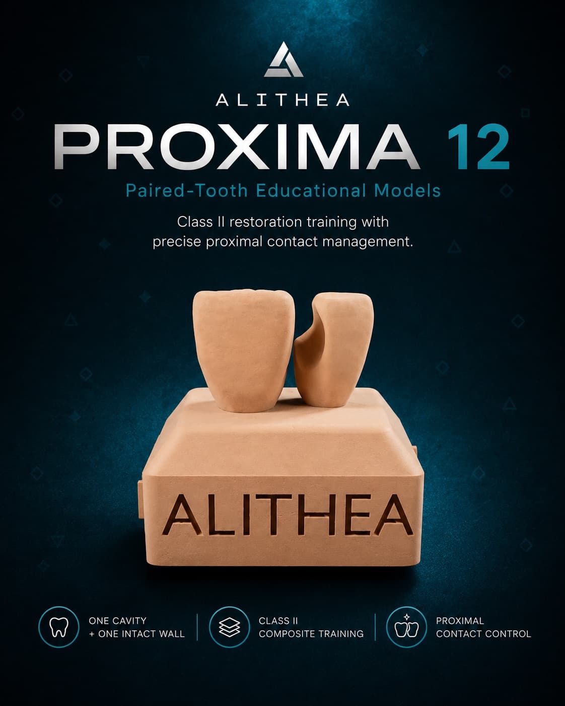
Proxima
Proximal restoration of the lateral incisor adjacent to the central to master the anterior junction.
MAD 20
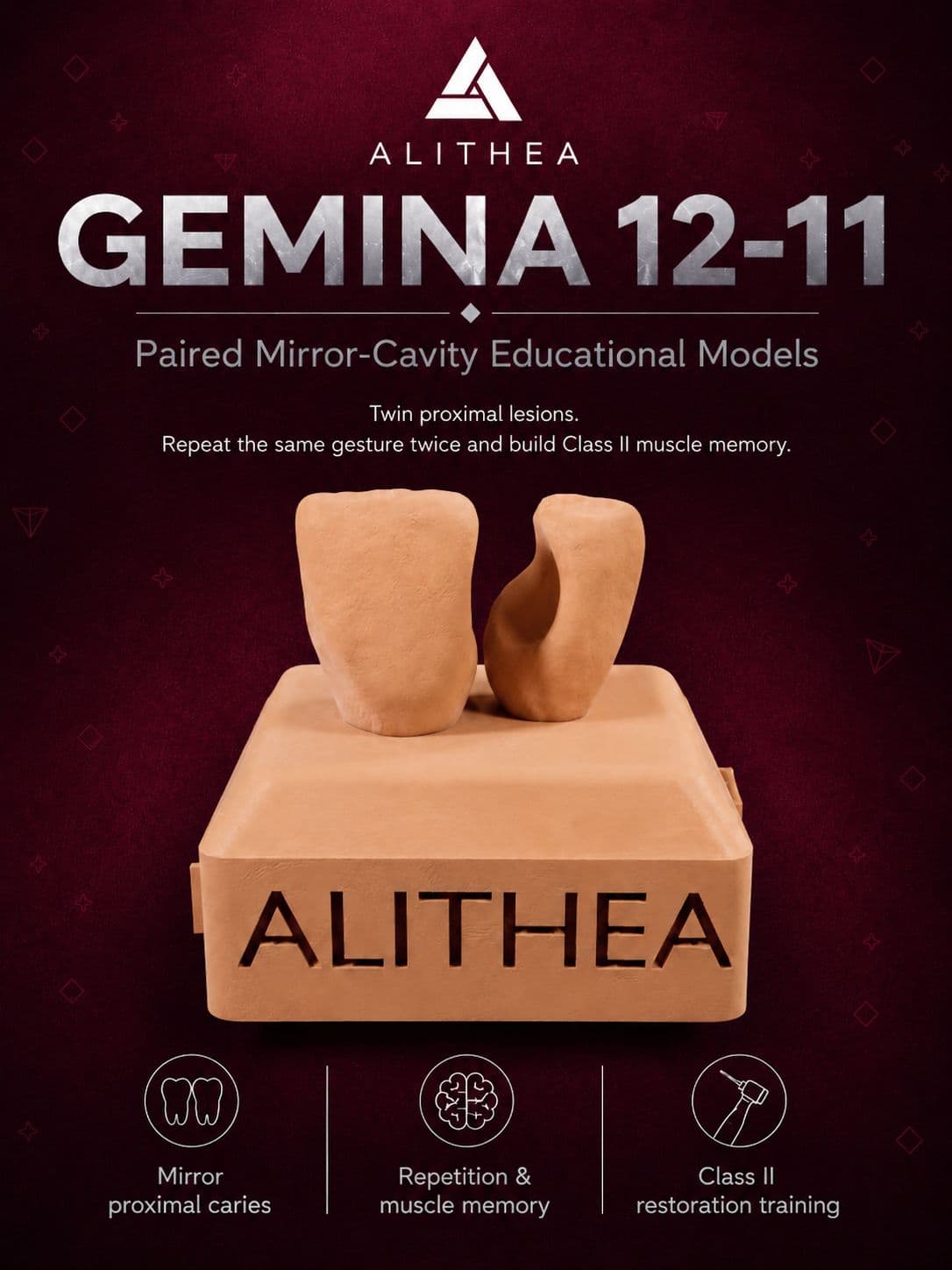
Gemina
Lateral and central incisor, each carrying a proximal lesion: the same gesture twice, in the most visible sector of the mouth.
MAD 20
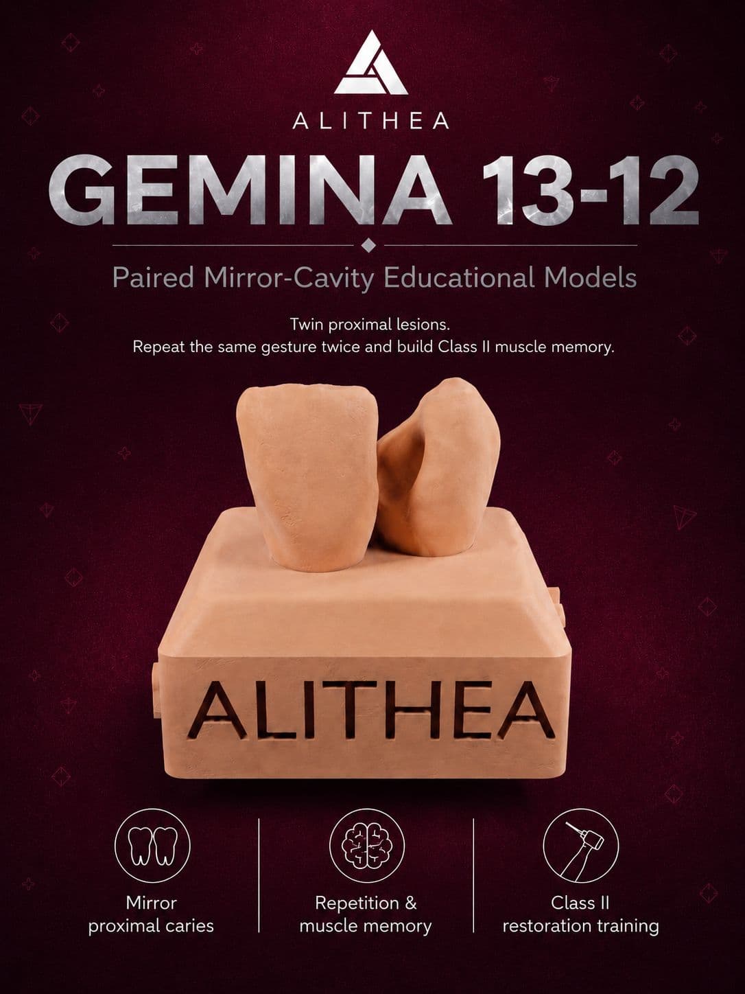
Gemina
Canine and lateral incisor, both affected — canine contact work with no intact reference left.
MAD 20
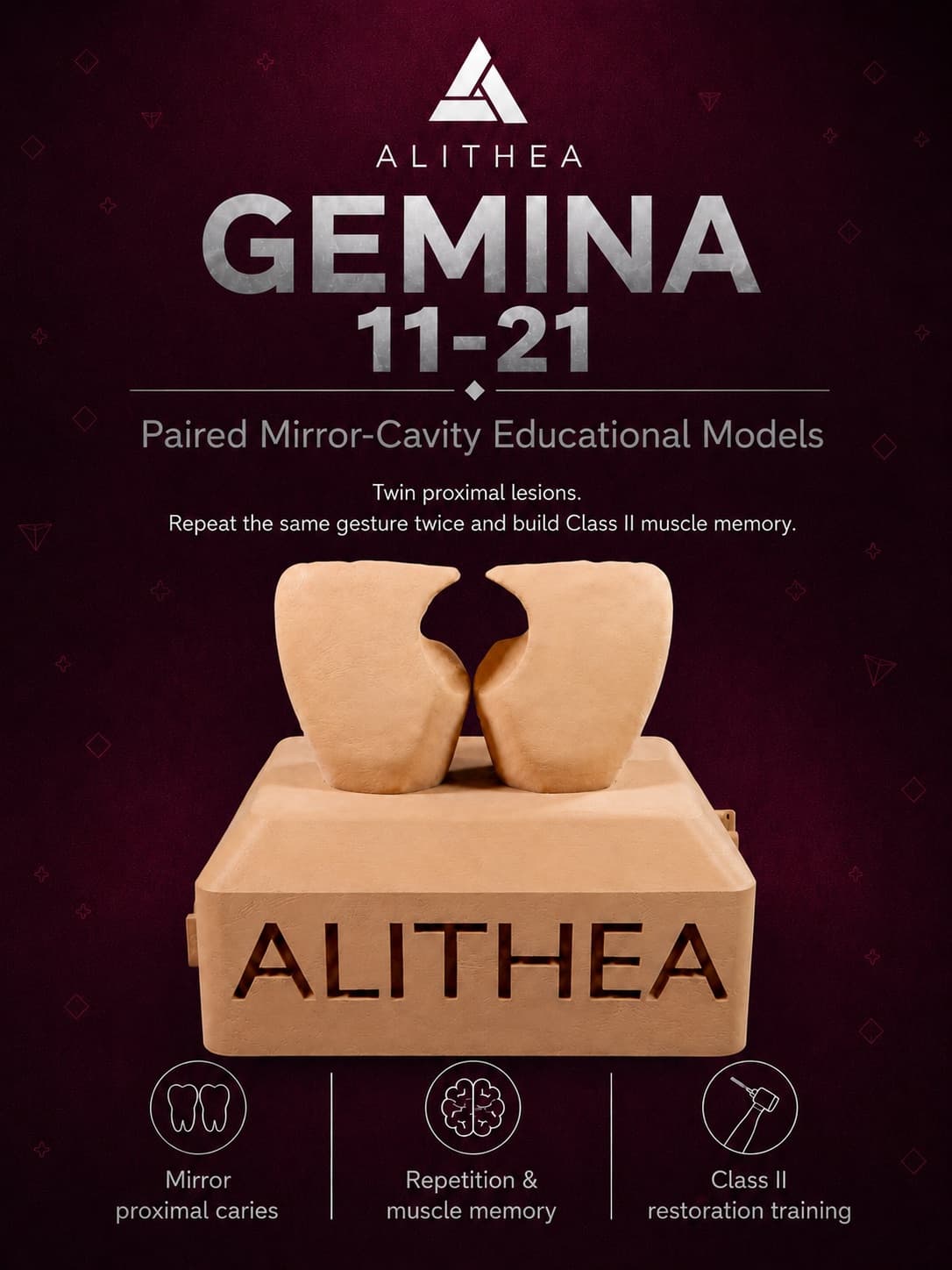
Gemina
Both central incisors with mirrored lesions: a tight contact to rebuild without shifting the midline.
MAD 20
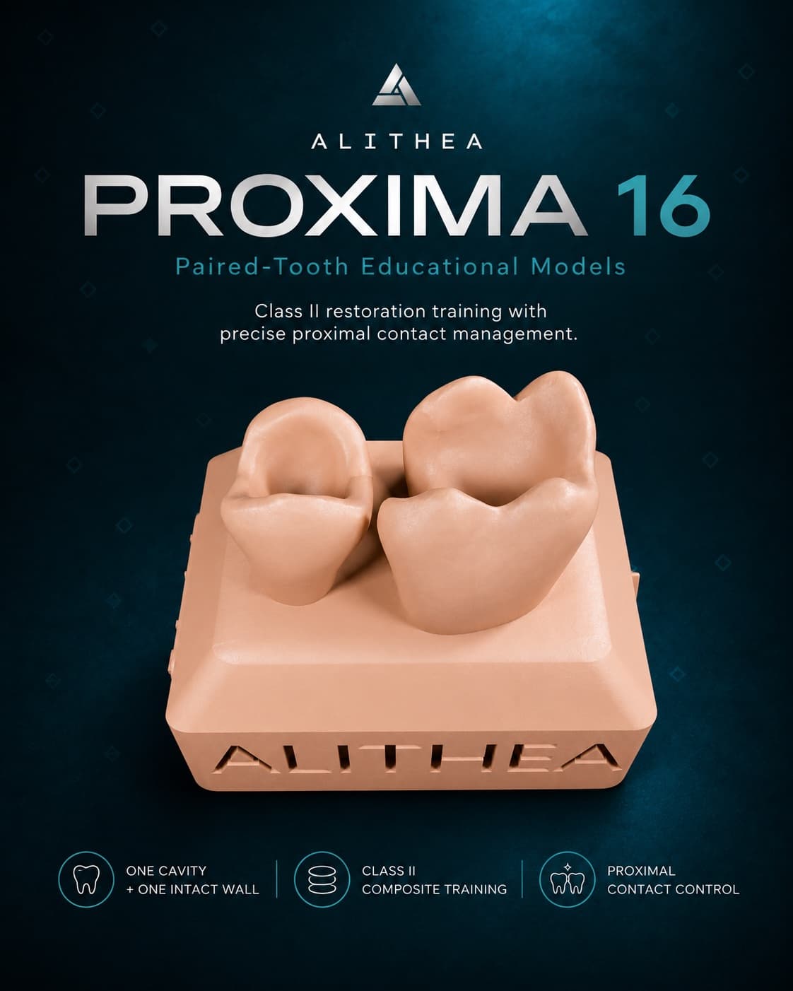
Proxima
Paired molar and premolar: a single Class II cavity on the 16, the neighbouring wall left intact to guide the contact.
MAD 20
Proxima
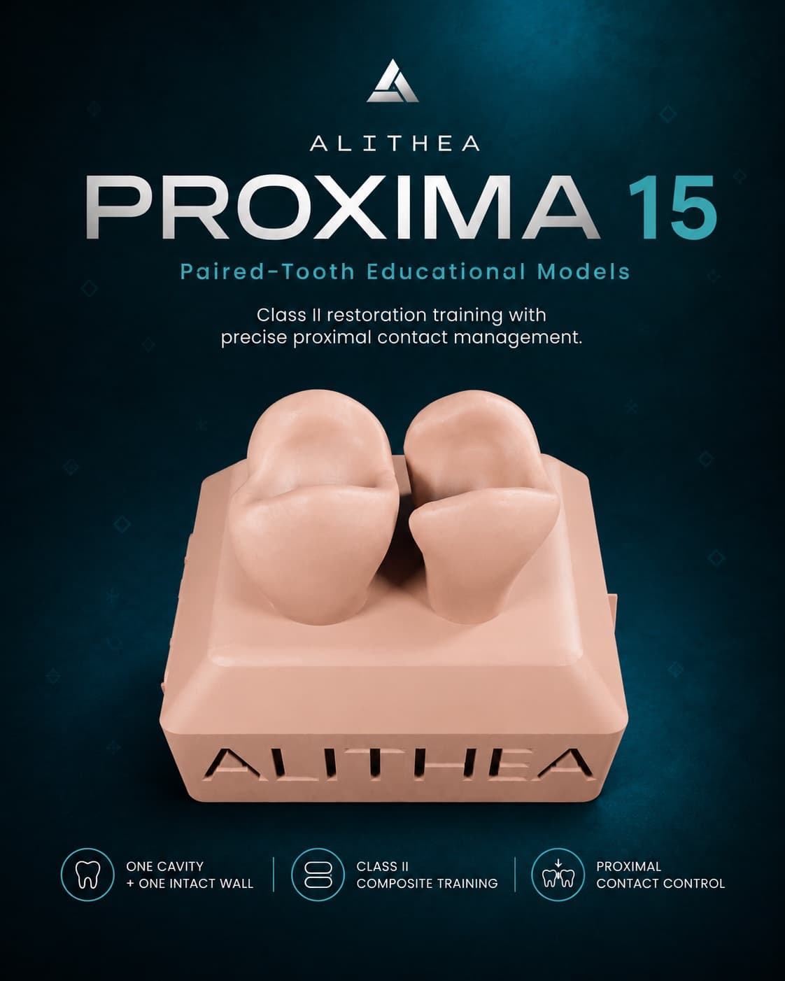
Maxillary premolars: one Class II cavity on the 15, the 14 left intact as the contact reference.
MAD 20
Proxima
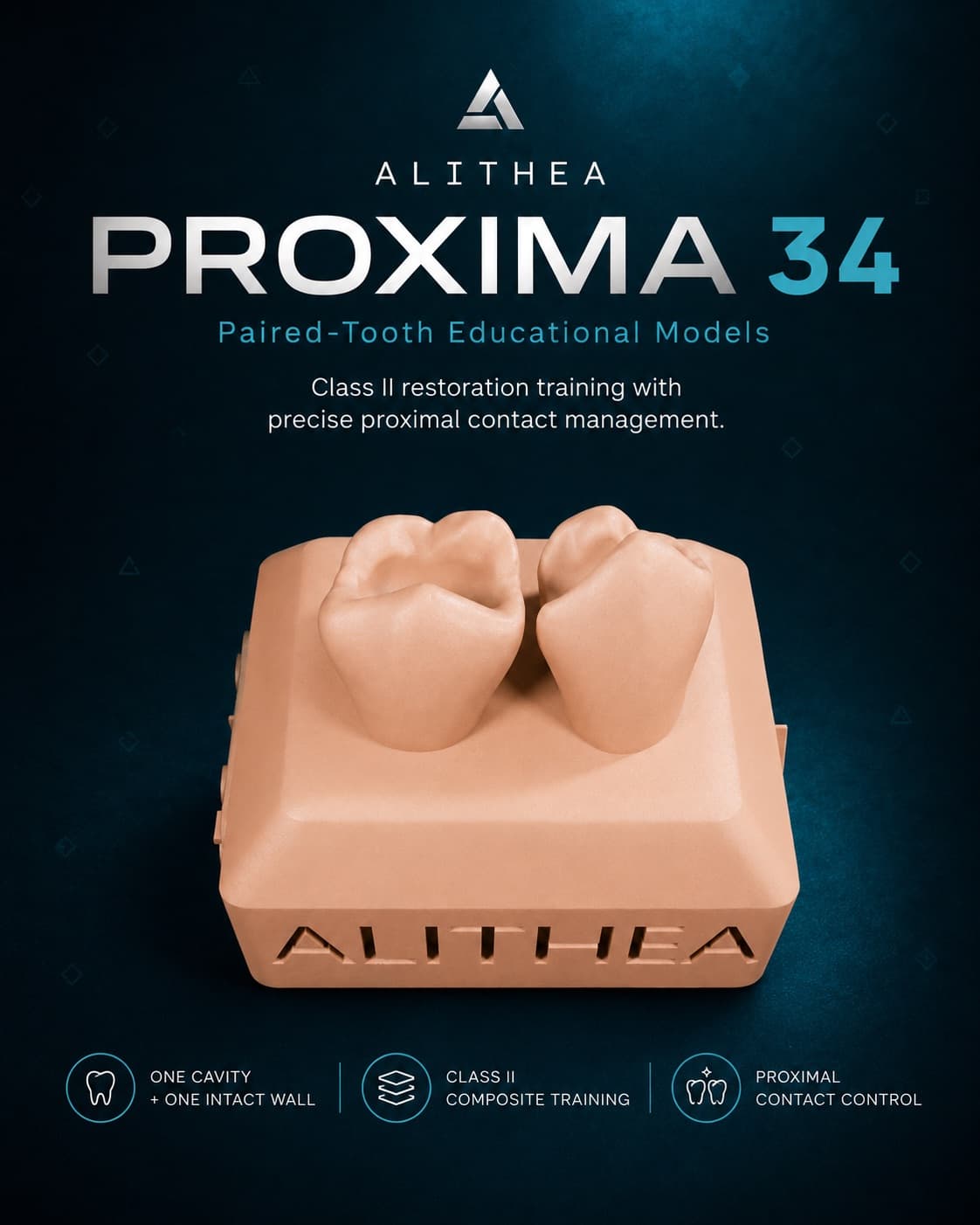
Mandibular premolars: one Class II cavity on the 34, the 35 intact to set the contact.
MAD 20
Gemina
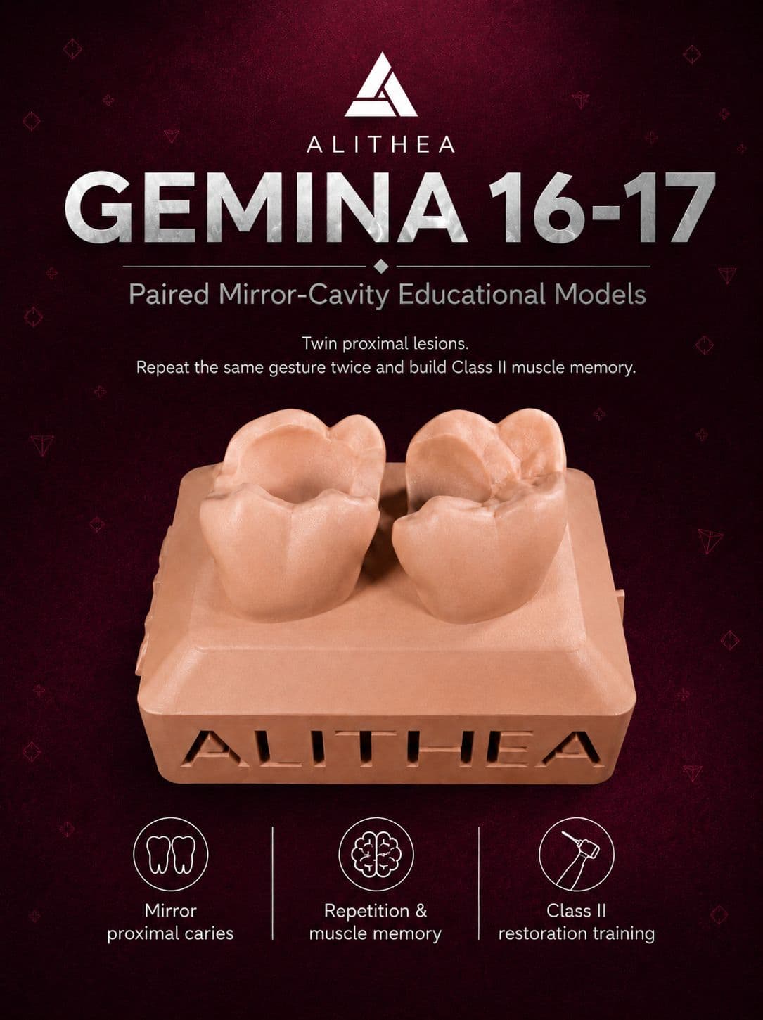
Two Class II lesions on adjacent lower molars for intensive repetition.
MAD 20
Gemina
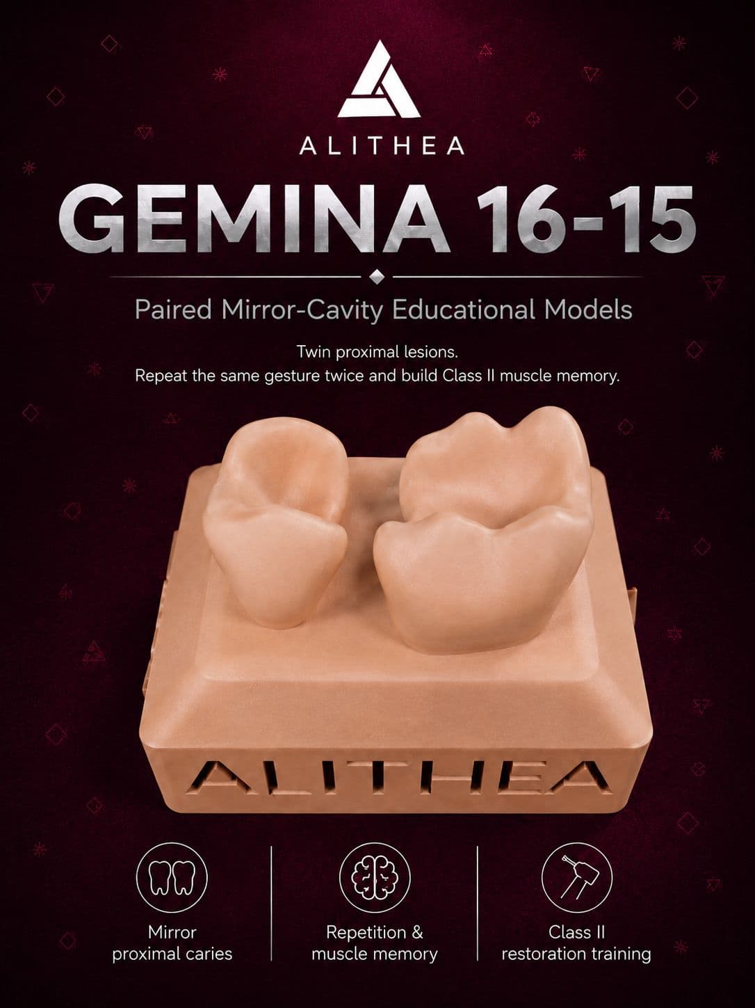
Extended Class II lesions on molar-premolar for training in complex restorations.
MAD 20
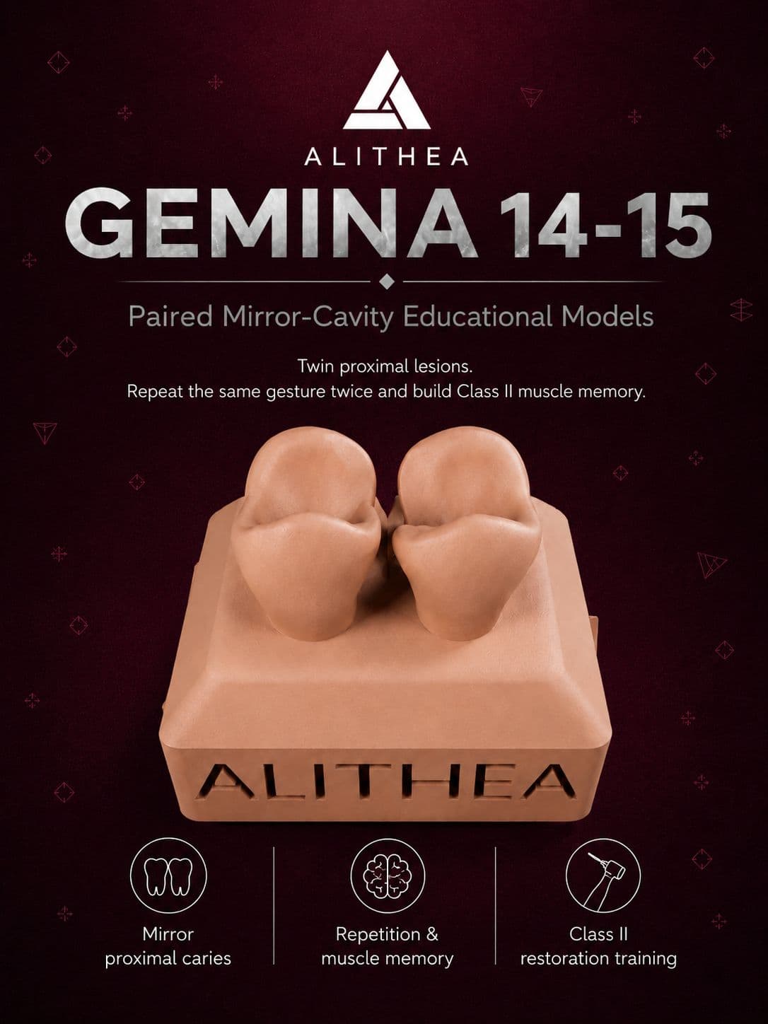
Gemina
Mesio-occluso-distal preparations on upper premolars for advanced training.
MAD 20
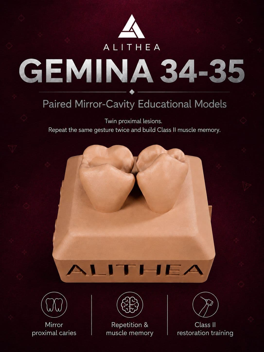
Gemina
MO-DO cavities on lower premolars for mastering mandibular biproximal restorations.
MAD 20
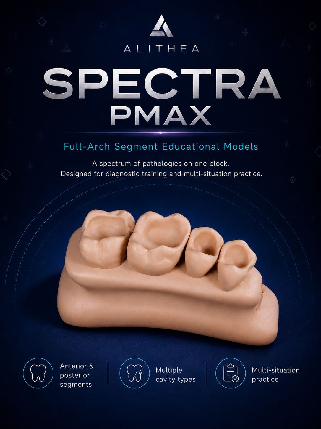
Spectra
Upper arch segment for multi-pathology reading and diagnostic training.
MAD 34
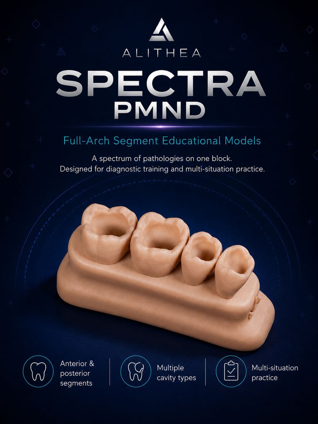
Spectra
Lower arch segment for sector diagnosis and treatment planning.
MAD 34
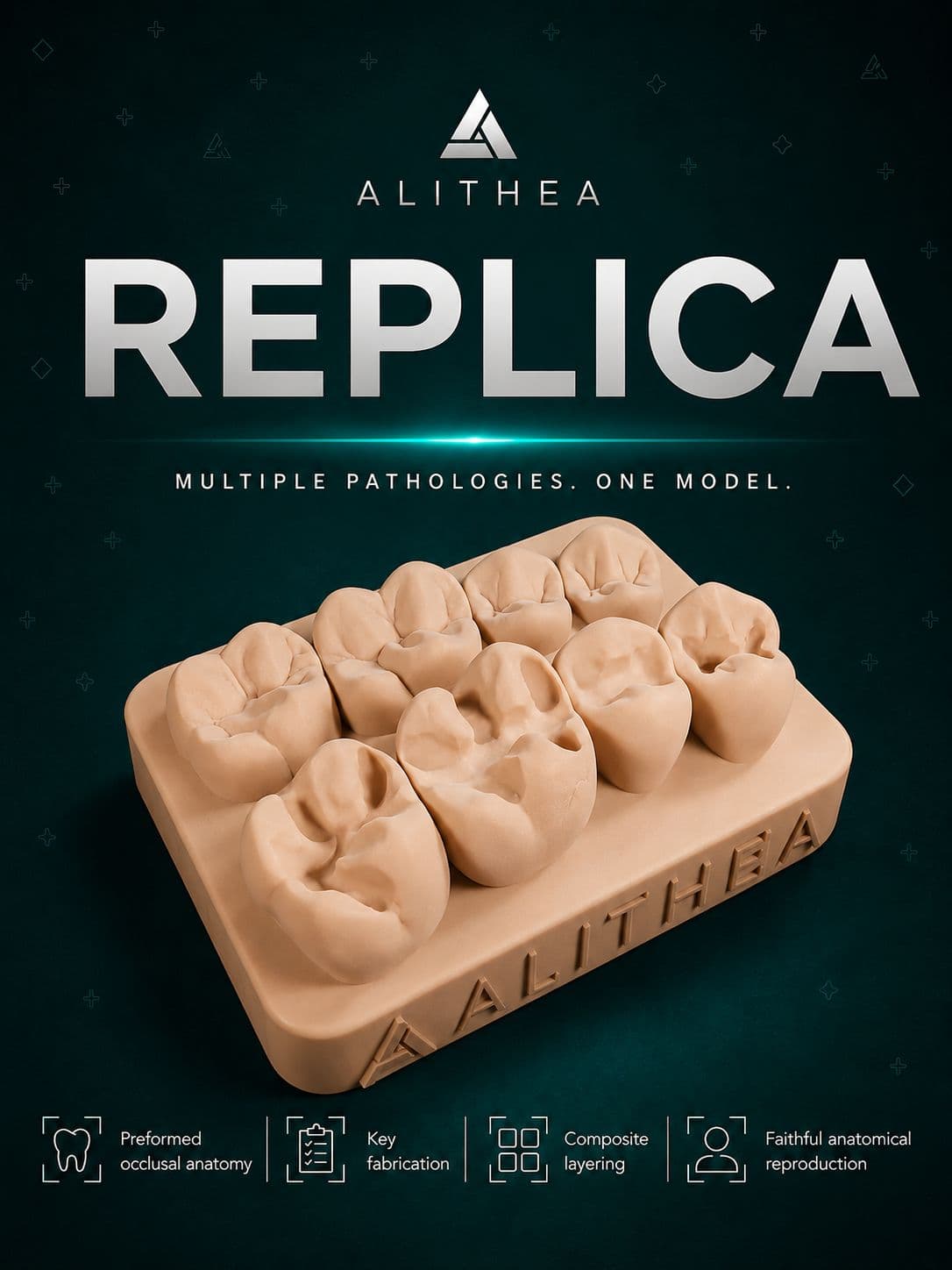
Replica
Preformed occlusal anatomy for learning the stamp technique and composite layering.
MAD 44